
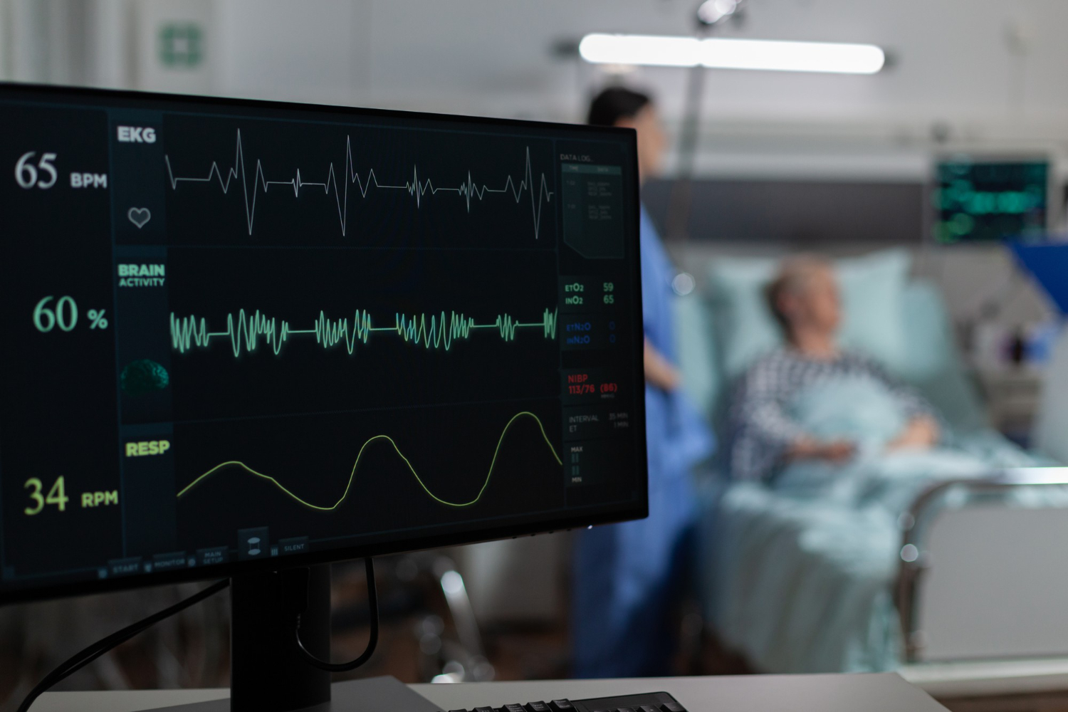
2D Echocardiography is a non-invasive diagnostic imaging test that uses ultrasound waves to examine the structure and functioning of the heart. It provides real-time images of the heart, helping healthcare professionals assess its chambers, valves, walls and pumping activity. 2D Echocardiography screening supports the clinical evaluation of cardiovascular health and helps identify abnormalities that may require further medical attention.
Heart health assessment is an important part of preventive healthcare, particularly for individuals experiencing symptoms such as chest discomfort, breathlessness, palpitations or unexplained fatigue. Echocardiography can provide valuable information about cardiac structure and function, supporting medical evaluation and appropriate treatment planning.
2D Echocardiography helps assess different aspects of heart function and structure, including:
A healthcare professional may recommend echocardiography based on symptoms, medical history or cardiovascular risk factors. The investigation may be useful for individuals experiencing:
2D Echocardiography is generally a painless and non-invasive procedure that does not involve radiation. During the test, a trained healthcare professional applies ultrasound gel to the chest and moves a handheld transducer over the examination area to obtain images of the heart.
The resulting images help assess cardiac anatomy and function. The findings are interpreted in the context of the patient’s symptoms and overall clinical condition.
2D Echocardiography screening supports cardiovascular evaluation by providing important information about heart structure and functioning. When clinically indicated, this investigation can assist in identifying cardiac abnormalities, monitoring existing conditions and guiding further medical management.
Early evaluation and appropriate cardiac assessment can help patients make informed decisions about their heart health and overall well-being.